27
May
This tutorial will demonstrate the lead placement for the 12 lead ecg of the limb leads (ra: The limb leads record the activity from a plane.

Where to put ecg leads. This modified ecg leads placement system suggests that the ecg limb leads could be placed on the left and the right acromial region for the upper limb leads, and on the left and right anterior superior iliac spine for the inferior limb leads. Next, apply lead 2 to the right arm. In the cabrera system, the leads are placed in their anatomical order.
In order to build up an electrical image of the electrical activity of the heart, you need to record from two orthogonal planes. I asked nurses, ekg technicians, medical assistance, and even cardiology fellows where ecg leads/electrodes should be placed on the patient’s body. Limb leads can be placed on any part of the patient’s respective limbs.
Again, the front of the shoulder is suggested here, in a place with little or no muscle or movement. When in doubt, repeat the ecg! The limb leads can also be placed on the upper arms and thighs.
Here is a detailed view of the pediatric 12 lead ecg placement approach: For instance, do not attach an electrode on the right wrist and one on the left upper arm. Leads are placed as shown:
The limb leads can also be placed on the upper arms and thighs. The limb electrodes should be placed on the right and left wrists and the right and left ankle. One may also ask, what is the difference between a 12 lead and 4 lead ecg?
This is the most useful lead for detecting cardiac arrhythmias as it lies close to the cardiac axis (the overall direction of electrical movement) and. The inferior limb leads (ii, avf and iii) are juxtaposed, and the same goes for the lateral limb leads and the chest leads. Then connect the left leg.
Most of the time the answer is “somewhere around here, and they point to areas on the arms, legs, and chest. Ecg leads (or electrocardiographic leads) record the electrical potential at the different points on the body. However, there should be uniformity in your placement.
There’s a reason for that. We suggest the front of the left shoulder in a place where there is little muscle or muscle movement, to avoid any emg signal disturbance. 12 lead ecg (ekg) placement of electrode stickers:
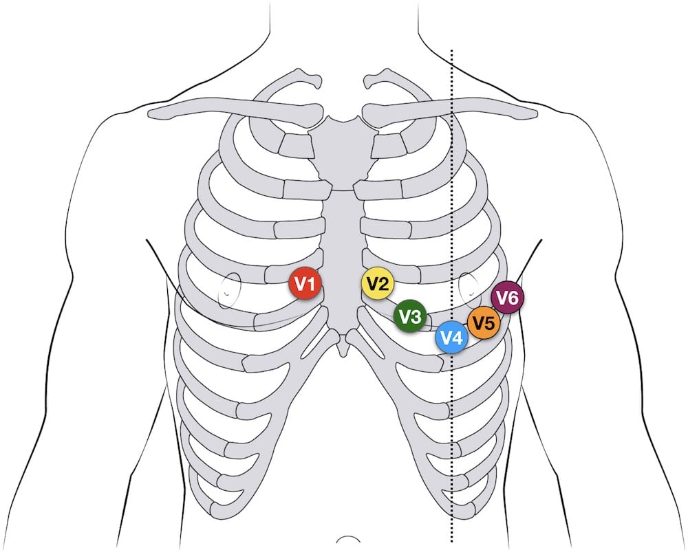
Right arm (ra) over the manubrium; Therefore, in female patients, the v4 , v5 and v6 leads are recommended to be placed underneath the left breast where the breast tissue meets the chest (qld doh 2012). The limb leads record the activity from a plane.
Place the lead at roughly the same location as you placed the lead on their right side to ensure that both arm leads give accurate readings. Breast tissue can impact on the ecg amplitude due to the increased distance between the electrode and the heart when ecg electrodes are placed over the chest (rautaharuju et al. If the patient has a defibrillator on their chest or has a pacemaker, do not place the ecg lead on the electrical device.
However, there should be uniformity in your placement. For instance, do not attach an electrode on the right wrist and one on the left upper arm. Your first clue is a negative qrs complex in lead i.
These leads are not suppose to go anywhere on the torso. Apply limb leads apply lead 1 to the left arm. In respect to this, where are the 12 leads placed on a patient for an ecg?
Arm leads are placed distal to the shoulder and legs leads distal to the hips. The most common is right and left arm reversals. Where the leads are placed determines a specific pattern we are looking for on our recording.
Press down firmly on the lead to fix it in place. This tutorial will demonstrate the lead placement for the 12 lead ecg of the limb leads (ra: The main ecg pointers for limb lead reversal:
Left arm (la) over the xiphoid process;
Previous post
Where was hiv first discoveredNext post
Where to get vaccines in nyc