16
Mar
Clinical implications, basis, lead configurations, and nursing implications Payment of a financialobligationearlier than is expected or required.

What are the precordial leads. (redirected from precordial leads) also found in: The sternal ridge/angle (“angle of louis”) is at the level of the 2 nd intercostal space. N engl j med 1999;
Additionally, inaccurate placement of precordial leads poses a It is a unipolar lead having two electrodes, one is positive or exploring electrode applied on the surface of the chest wall and other is indifferent electrode. Copyright © 2012, campbell r.
As a result of the location of the ground lead (wct) being in the center of the chest, the precordial leads have exploring electrodes located anteriorly on the chest wall. The standard six precordial leads are well selected in that they span the anterior surface of the heart from precordial position 1, which overlies the right auricle, to position 6, which overlies the anterolateral portion of the left ventricle. It is an electrocardiographic manifestation of a critical proximal left anterior descending coronary artery stenosis in patients with unstable angina and is characterized by symmetrical, often deep t wave inversions in the anterior precordial leads, little or no cardiac marker elevation, discrete or no st segment elevation and no loss of precordial r waves [6].
The precordial (chest leads) leads each consist of a positive electrode strategically placed on the chest of the patient. There are six precordial leads. They are unipolar leads, they register the absolute potential of the point where the electrode of the same name is placed.
Hence, the chest leads are excellent for detecting vectors traveling in the horizontal plane. Lead misplacement has previously been shown to cause changes on the ecg with shifts of precordial leads by 2 cm resulting in altered r wave progression and shift in the precordial transition zone in 20% and 75% of patients, respectively ,. Low voltage may be present in the following situations:
Ecg, electrocardiograph, unipolar leads precordial lead a lead having one electrode placed over the precordium, the other over an indifferent region. In addition to the three standard limb leads and the three augmented limb leads that view the electrical activity of the heart from the frontal plane, there are six precordial, unipolar chest leads. Contrastingly, precordial leads are bundled together as a group.
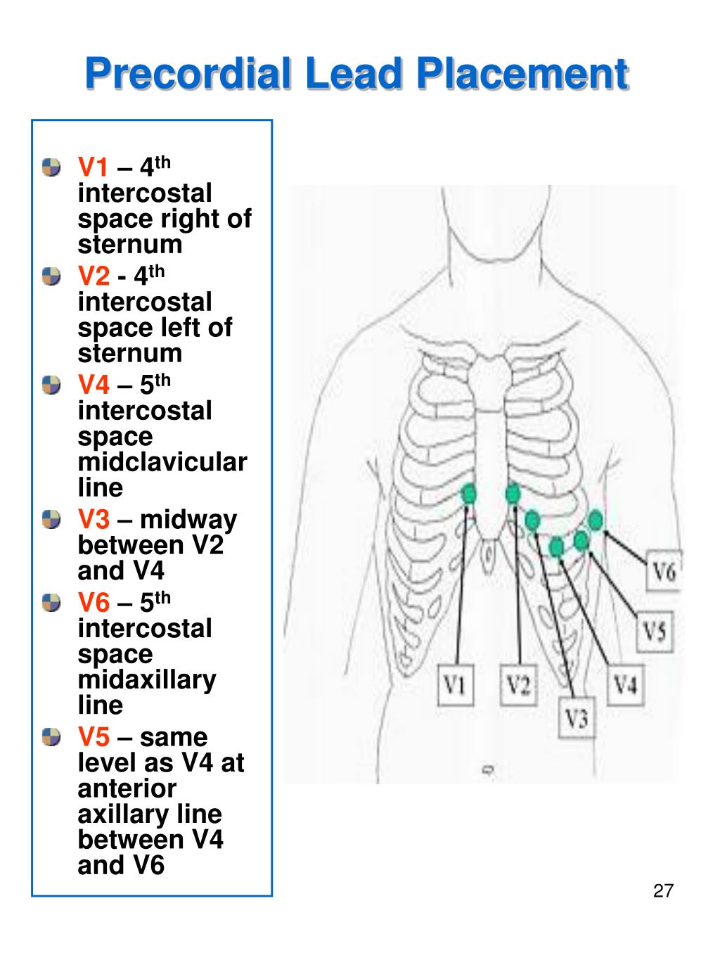
This configuration places six positive electrodes on the surface of the chest over different regions of the heart in order to record electrical activity in a plane perpendicular to the frontal plane. A precordial lead is one of the leads placed on the chest for an electrocardiogram. The positions of the positive electrode for the six precordial leads are very important for a valid tracing to be made on the ekg machine.
The remaining six leads (designated as leads v1, v2, v3, v4, v5, and v6) are the chest or precordial leads (wilson et al., 1944). The right precordial leads (v1, v2 and v3) are critically important in diagnosing posterior wall stemis. They are designated by a capital v and a number from 1 to 6.
They are the leads best suited for pinning down left ventricle abnormalities, especially of the anterior and posterior walls. Payment of a financialobligationearlier than is expected or required. Clinical implications, basis, lead configurations, and nursing implications
The anatomic position of the septum is well correlated with the position of the electrical. The value of the right precordial leads of the electrocardiogram. The chest (precordial) leads (v1, v2, v3, v4, v5 and v6) have the exploring electrodes located anteriorly on the chest wall and the reference point located inside the chest.
Previous post
What are the treatments for osteoporosisNext post
What are the options