
08
Mar
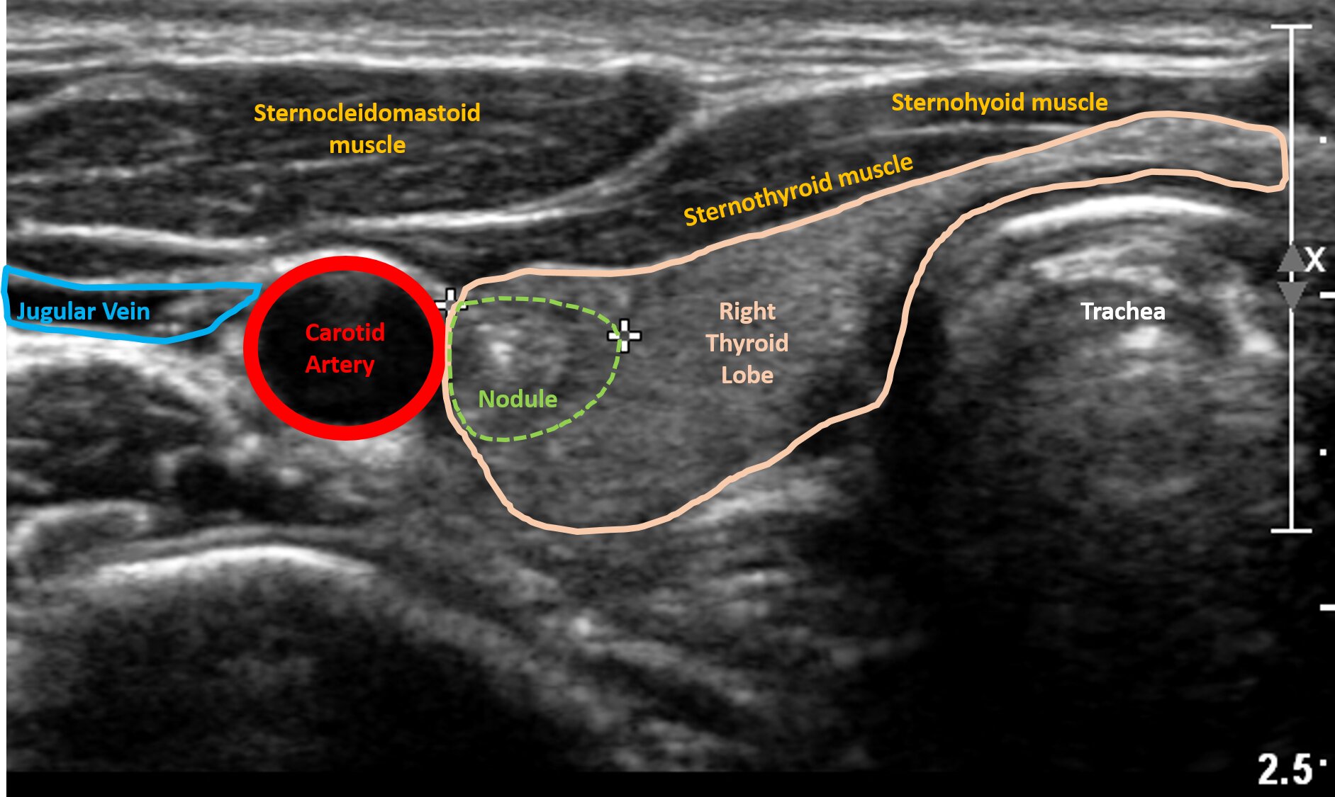
It measures 0.57 x 0.61 x 0.69cm. The thyroid gland is located in front of the neck just above the collar bones and is shaped like a butterfly, with one lobe on either side of the neck connected by a.
Ultrasound images of thyroid cancer. Browse 127 thyroid ultrasound stock photos and images available, or search for thyroid ultrasound mask to find more great stock photos and pictures. In ultrasound images, the cancer regions are usually blurred, vague margin and irregular in shape. Colour doppler examination shows mild increased vascularity.
Ultrasound showed a grossly enlarged left thyroid lobe measuring 8.5cm in length and 5.6cm in width. Fine debris is present in parts of the cystic mass which is seen to occupy most of the left lobe of thyroid. They can also help determine whether, and how far, the cancer may have spread.
Neck ultrasonography is the best, most sensitive and specific method of evaluating thyroid nodules, thyroid goiters, and neck lymph nodes. An ultrasound of the thyroid produces pictures of the thyroid gland and the adjacent structures in the neck. See more ideas about thyroid ultrasound, ultrasound, thyroid.
The ultrasound images of the thyroid cyst show the lesion filled with clear fluid with some septation towards the posterior half of the hrmorrhagic cyst. She was referred to government hospital and a ct scan was done. Ultrasound uses soundwaves to create a picture of the structure of the thyroid gland and accurately identify and characterize nodules within the thyroid.
Ultrasound is critical in detection, diagnosis, and management of thyroid nodules. An improved deep learning approach for detection of thyroid papillary cancer in ultrasound images. Ultrasound (us) has facilitated the qualitative diagnosis of thyroid nodules, however, some mtc may be diagnosed as a benign nodule on ultrasound because ultrasound features of malignancy are lacking.
To help raise awareness, we wanted to show you photos of people living with thyroid cancer as well as thyroid cancer survivors. The aim of the study was to investigate the association between ultrasound. The two scans are identical, the one on the right is outlined to help you understand what you are looking at.
It measures 0.57 x 0.61 x 0.69cm. The above image is an ultrasound of a typical thyroid nodule, except that this nodule is a bit bigger than usual. Ultrasound images & clips papillary thyroid carcinoma with an ill defined inhomogeneous mass with calcifications in the right thyroid lobe and lymph node metastasis papillary thyroid carcinoma with with calcifications longitudinal
Ultrasound images & clips anaplastic thyroid carcinoma with an irregular hypoechoic mass with calcifications in in the left lobe with lymph node metastases The mass is solid with heterogenous echotexture. Color doppler showed increased vascularity, while the rest of the thyroid showed normal vascularity.
Specifically, we highlight morphologic characteristic of the nodule, including its echo signal in relation to its consistency, nodular size, number and contour. Ultrasound is also frequently used to guide the needle into a nodule during a thyroid nodule biopsy. The thyroid gland is located in front of the neck just above the collar bones and is shaped like a butterfly, with one lobe on either side of the neck connected by a.
Medullary thyroid cancer (mtc) has more aggressive behavior and poor prognosis. If your doctor suspects that you have thyroid cancer, they may order a variety of imaging tests,. The use of ultrasound in thyroid cancer imaging is dealt with in more detail in the later part of the review, but in brief, its major roles include:
A common imaging test used to evaluate the structure of the thyroid gland.
Previous post
University of arlington texas rn to bsnNext post
Ultrasound guided injections hip