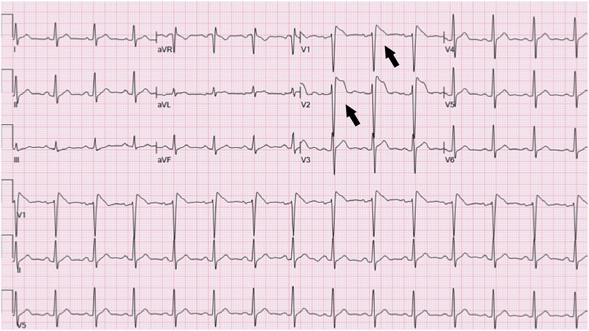
In the case of subendocardial ischaemia, st elevation in avr is simply a reciprocal change to st depression in these leads. The st segment is concave.
Coronary athersclerosis and presence of high risk thin cap fibroatheroma (tcfa) can result in.
St elevation in ecg. St segment on an ecg can be elevated for a number of reasons: St elevation in avr ddx. Ecg 1 h after initiation of treatment.
Most of the st depression patterns seen during st elevation myocardial infarction represent reciprocal changes rather than ischaemia at a distance. Dominant r wave in v1/2 (like right ventricular hypertrophy but without other changes), st depression. It is associated with extensive myocardial damage and paradoxical movement of the left ventricular wall during systole.
The st segment is concave. In the setting of chest discomfort (or other symptoms suggestive of myocardial ischemia) st segment elevation is an alarming finding as it indicates that the ischemia is extensive and the risk of malignant arrhythmias is high. St elevation during stress ecg is an abnormal and fairly unusual response.
Chew school of medical sciences universiti sains malaysia. Coronary athersclerosis and presence of high risk thin cap fibroatheroma (tcfa) can result in. St depression does not localise, and thus subendocardial ischaemia due to oxygen supply/demand mismatch produces a consistent ecg pattern of lateral st depression and reciprocal st elevation in avr.
St segment elevations in ecg. The characteristic ecg changes consistent with stemi are: Most commonly it is seen in the presence of pathologic q waves, where it may suggest an lv aneurysm or periinfarction ischemia.
Introduction st segment of the cardiac cycle represents the period between depolarization and repolarization of the left ventricle in normal state, st segment is isoelectric relative to pr.</p> Minimally peaked t waves (arrows) and sinus tachycardia. 82 however, one ecg pattern, st depression in leads v5 and v6 in acute inferior myocardial infarction, does signify concomitant coronary artery disease of the lad vessel with acute ischaemia in a.
In the case of subendocardial ischaemia, st elevation in avr is simply a reciprocal change to st depression in these leads. St elevation in >2 small squares in 2 adjacent chest leads, st elevation > 1 small square in 2 adjacent limb leads, or new lbbb ischaemia: Pericarditis is associated with ecg.
Serum potassium measured 9.4 mmol/l. St segment elevations in ecg k.s. Remember, these patients typically look sick and are actively.
