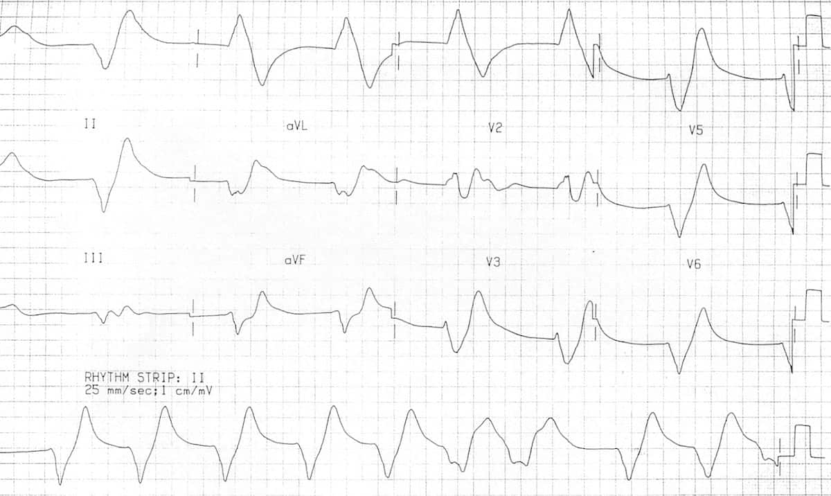
Tall, peaked t waves with a narrow base (best seen in precordial leads) progressive flattening and eventually disappearance of p waves; Virginia scarabeo, maria stella baccillieri, attilio di marco, fabio de conti, francesco contessotto, piergiuseppe piovesana journal of cardiovascular medicine 2007, 8 (9):

Hyperkalemia can present with multiple abnormalities on an ecg, including.
Sine wave pattern ecg. Is sound a sine wave? In 1972, manseau et al. Hyperkalaemia is defined as a serum potassium level of > 5.2 mmol/l.
Severe hyperkalem ia may be devastating to cause. Ekg or ecg waveform parts are explained clearly to make ekg interpretation easy. The earliest manifestation of hyperkalaemia is an increase in t wave amplitude.
2 furthermore, rapidly progressing flaccid motor weakness may result in a quadriplegia, which was the presenting symptom in this case and is an uncommon. Tall, peaked t waves with a narrow base (best seen in precordial leads) progressive flattening and eventually disappearance of p waves; Absence of the p waves and, eventually, a sine wave pattern (see below), which is frequently a fatal rhythm.
Enlarge giving iv calcium is cardioprotective in the setting of hyperkalemia. A sine wave or sinusoid is any of certain mathematical curves that describe a smooth periodic oscillation. Abnormal the new nichd guidelines label four fhr patterns as abnormal.
I don�t know if this was a pacemaker provided in the ed, or if the patient has an implanted pacer. Hannibal is staff nurse, progressive care unit, the louis stokes cleveland department of veterans affairs medical center, 10701 east boulevard, cleveland, oh 44106 ( jerry.hannibal@gmail.com ). Sine wave elect rocardiogram (ecg) pattern in hy perkalemia is still a m edical curiosity and rare to observe in routine clinical practice.
To address the clinical significance of sinusoidal heart rate (shr) pattern and review its occurrence, define its characteristics, and explain its physiopathology. The morphology of this sinusoidal pattern on ecg results from the fusion of wide qrs complexes with t waves. Provides information on atrial depolarization and the p wave, ventricular depolarization a
The qrs complex becomes wider. Virginia scarabeo, maria stella baccillieri, attilio di marco, fabio de conti, francesco contessotto, piergiuseppe piovesana journal of cardiovascular medicine 2007, 8 (9): It occurs often in both pure and applied mathematics, as well as physics, engineering, signal processing and many other fields.
Ecg changes generally do not manifest until there is a moderate degree of hyperkalaemia (≥ 6.0 mmol/l). What category is a sinusoidal pattern? But the levels at which ecg changes are seen are quite.
Plasma ph was normal and flecainide drug levels were initially unavailable. There is a sine wave, which is seen with severe hyperkalemia.it is sometimes called ventricular flutter, and is also reported with class i antidysrhythmics, which are sodium channel blocking agents.there are pacer spikes. As the severity of hyperkalemia increases, the qrs complex widens and the merging together of the widened qrs complex with the t wave produces the ‘sine wave’ pattern of severe hyperkalemia.
The sine wave pattern depicts worsening cardiac conduction delay caused by the elevated level of extracellular potassium. Its most basic form as a function of time is: Y = a sin .
Severe hyperkalemia with sine wave ecg pattern gerard b. Johnson francis | december 2, 2014. Previously mentioned ecg changes become more pronounced.
This is just one of the 7 puzzles found on today’s bonus puzzles. Hyperkalemia can present with multiple abnormalities on an ecg, including. It is named after the function sine, of which it is the graph.
An ecg on arrival (figure 1) demonstrated an irregular broad complex bradyarrhythmia with a single p wave. Welcome to the page with the answer to the clue sine wave pattern. You can make another search to find the answers to the other puzzles, or just go to the homepage of 7 little words.
A sine wave is a continuous wave. Qrs widening is due to the reduced vmax as the intracellular negativity. September 19, 2020 mysticwords daily, seven.
Hannibal, rn, msn, pccn gerard b. Tall peaked t waves in hyperkalemia is due to the enhanced outward potassium current ikr. The classical ecg change in hyperkalemia is tall tented t waves.
This is certainly alarming because sine wave pattern usually precedes ventricular fibrillation.