
02
Nov
Tibial pain scores were not predictive of stress fracture. The main cause of bone stress injuries, including tibial stress fractures and medial tibial stress syndrome, is increased training loads coupled with inadequate rest.
Medial tibial stress fracture. Many have advocated the term medial tibial stress syndrome to refer to anterior shin pain as a result of exercise. Rest should be included in the training planning as an equally important part of the training regime as the training itself. So, this refers to the location of the stress fracture.
We found that mri showed three different localizations of medial tibial plateau stress fractures, which were associated with tibial posterior slope at the medial tibial plateau. The main cause of bone stress injuries, including tibial stress fractures and medial tibial stress syndrome, is increased training loads coupled with inadequate rest. 14 the term shin splints traditionally has been used synonymously with mtss.
Medial tibial stress syndrome (mtss) is a common overuse injuries of the lower extremity, often seen in athletes and military personnel. Tibial pain scores were not predictive of stress fracture. Symptoms of a stress fracture in the posteromedial tibial border are similar to that of medial tibial stress syndrome (mtss) (commonly known as «shin splints»);
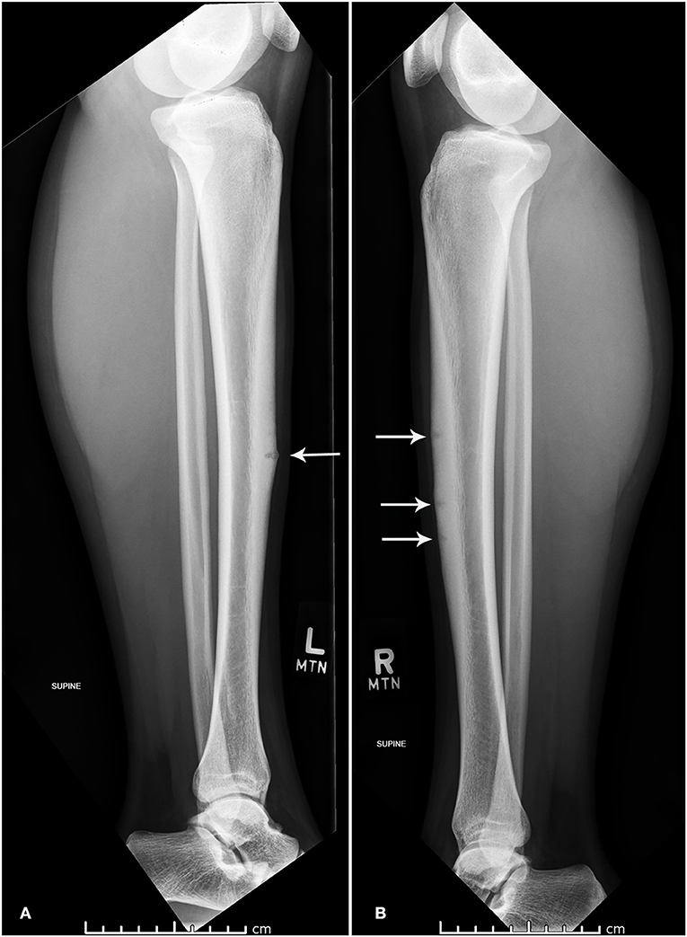
This type of stress fracture is rather rare. The tibial plateau is the top end of the bone at the knee joint. Tibial pain scores were not predictive of stress fracture.
A medial tibial plateau stress fracture would be on the side of the bone that faces the other leg. Tibial stress fractures are small cracks in the cortex of the bone which are usually due to overuse and repetitive stress, such as due to long distance running. Medial tibial stress syndrome (shin splints) can be distinguished from tibial stress fractures by diffuse tenderness along the length of the.
Medial tibial stress syndrome (mtss), a common overuse syndrome, is a periostitis or stress reaction characterized by diffuse pain along the posteromedial border of the tibia and associated with the tendon of the soleus. A tibial stress fracture is a condition that is primarily characterised by an incomplete break in the lower leg / shin bone (tibia) (figure 1). However, this term is ill defined and can.
[3][13] [14] however, periostitis, medial tibial stress syndrome (mtss) and tibial stress fracture should be viewed as a continuum of. This validation study provides the clinician with evidence based guidelines for the clinical diagnosis and treatment of medial stress fractures and their differentiation from shin splints. All multiple focal areas of signal abnormality in grade 4a stress injuries were located in the anterior and posterior tibial cortex (figs.
Medial tibial plateau morphology and stress fracture location: (also known as stress fracture of the tibia, medial tibial stress fracture) what is a tibial stress fracture? Crural fascia and muscle origins related to medial tibial stress syndrome symptom location.
A magnetic resonance imaging study. Stress fractures tend to occur as a result of overuse and are known as overuse injuries. Yates, b., & white, s.
Although stress fractures of the tibial diaphysis are common among athletes, the proximal tibial metaphysis is an unusual location for such injuries. Stress fracture of the tibia refers to a fatigue injury of the bone as a result of repetitive loading that overwhelms its capacity to heal and must be differentiated from medial tibial stress syndrome, which is not a stress fracture. Mri scan has proved very sensitive for diagnosing stress fractures and provided a definite diagnosis in our case.
Medial tibial stress fracture was found to occur when the band of tibial tenderness was ≤10cm in length. A stress fracture is a type of incomplete fracture in a bone. Medial tibial stress fracture was found to occur when the band of tibial tenderness was ≤10 cm in length.
Periosteal edema most commonly involved the posterior tibial cortex for grade 4b stress injuries and the medial tibial cortex for the remaining grades of stress injury.
Previous post
Mediastinal large b cell lymphomaNext post
Medial epicondyle fracture treatment