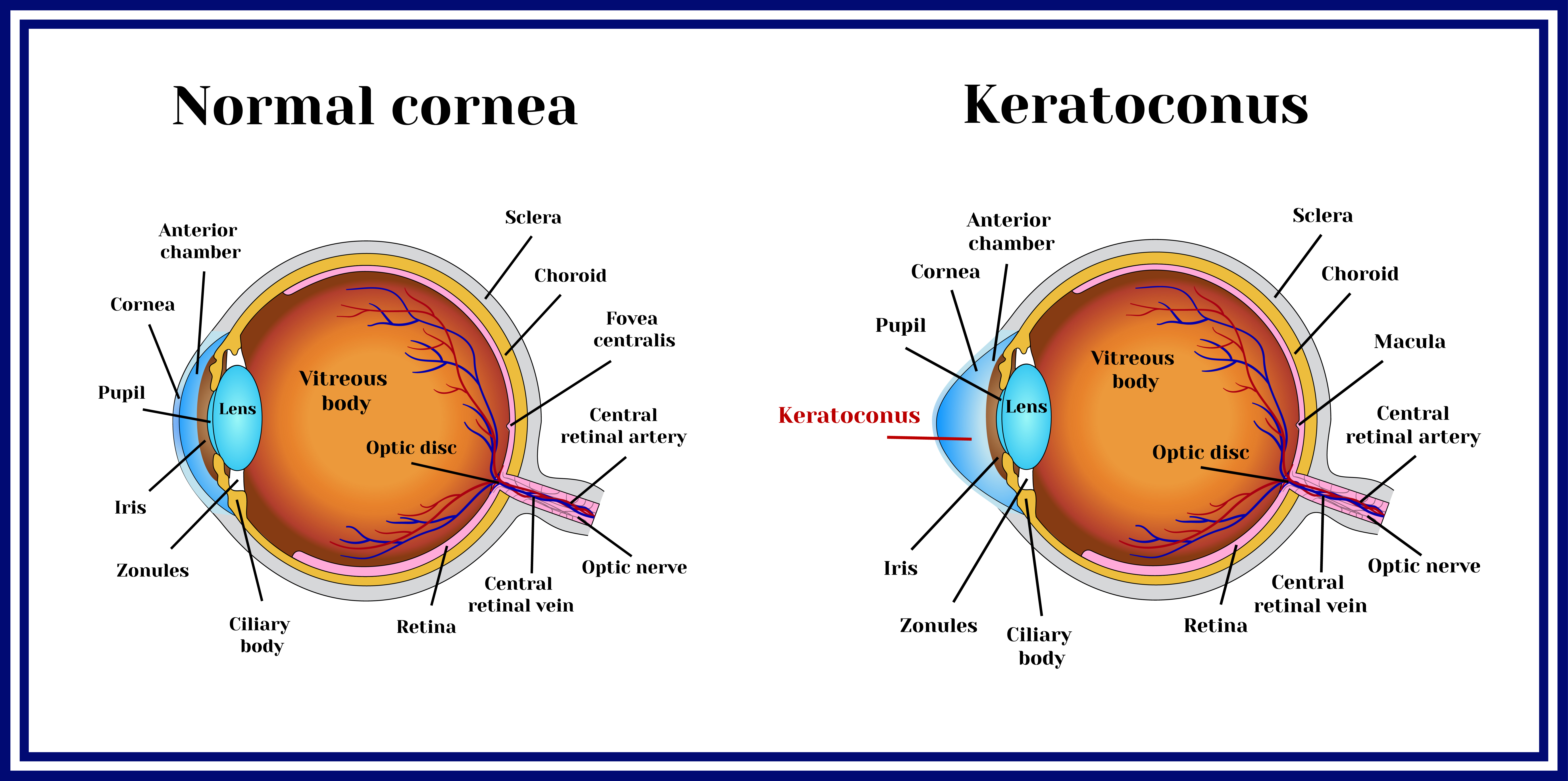
In keratoconus, the cornea (the front clear window of the eye) can become weak, thin, and irregularly shaped. Any of the following changes have occurred within 24 months:

You may, however, need glasses or contact lenses after the surgery for improved eyesight.
Corneal cross linking for keratoconus. This approval only covers the crosslinking products developed by glaukos corporation (formerly avedro, inc.). It is successful in more than 90% of cases. In this condition, the front part of your eye, called the cornea, thins out and gets weaker over time.
As we know, keratoconus occurs when the fibers that usually hold the cornea in an evenly domed shape become weaker, making them less effective at holding the cornea in position. Corneal cross linking (cxl) is a procedure to strengthen the cornea and stabilize the corneal changes caused by keratoconus or other corneal disease. Historically, as many as 1 in 5 patients with progressive keratoconus have required a corneal transplant, with more than half needing multiple transplants within 20.
Keratoconus is a condition of conical corneal shape where an irregularly shaped cornea prevents light from focusing correctly on the retina at the back of the eye. Cxl treatment is successful in more than 90% of cases. In keratoconus, the cornea (the front clear window of the eye) can become weak, thin, and irregularly shaped.
Corneal cross linking for keratoconus. Fda has approved corneal collagen crosslinking (cxl) for progressive keratoconus in april 2016. Corneal cross linking surgery for keratoconus stabilization corneal collagen cross linking is a process of strengthening and increasing the molecular bridges that hold the corneal layers together, therefore decreasing the chances of a bulge.
Using a variety of prescribed treatments, many patients have experienced improved quality and quantity of vision. The minimally invasive treatment involves applying liquid. Instead of keeping its normal round shape, corneas with keratoconus can bulge forward into the shape of a cone.
Any of the following changes have occurred within 24 months: Keratoconus as the most common cause of ectasia is one of the leading cause of corneal transplants worldwide. As a result, the cornea begins to bulge outwards in a conical shape.
However, several ocular and systemic associations exist like. Doctors use vitamins and light to amend unusual eye shape. It cannot reverse the disease, but it can slow it or stop it from progressing further and preserve your remaining vision.
According to the systematic review, cxl may be effective in halting the progression of keratoconus for one year under certain conditions, although evidence is limited due to the significant heterogeneity and paucity of rcts. The current available therapies do not modify the underlying pathogenesis of the disease, and none of the available approaches but corneal transplant hinder the ongoing ectasia. You may, however, need glasses or contact lenses after the surgery for improved eyesight.
Diagnosis of keratoconus based on keratometry and corneal mapping; After treatment, you will still need to wear spectacles or contact lenses.