Read more 5.3k views reviewed >2 years ago They can also show chronic lung conditions, such as.

The data set is organized into 3 folders (train, test, val) and contains sub folders for each image category (pneumonia/normal).
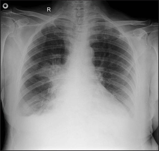
Chest x ray showing pneumonia. Pittii with a large pulmonary cavity of the right lower lobe, at hospital admission from publication. The data set is organized into 3 folders (train, test, val) and contains sub folders for each image category (pneumonia/normal). In its more common manifestation, pneumonia is caused by a bug that forms pus in the airways and alveoli, resulting in consolidation in part of the lung.
In other words, you have a white blob on your chest radiograph that indicates a pneumonia. Browse 64 chest x ray showing pneumonia stock photos and images available, or start a new search to explore more stock photos and images. Lobar pneumonias have specific appearances, which can be explained by the anatomical relationships of the lobes of.
Chest x ray pneumonia vs bronchitis is a general term in widespread use, defined as infection within the lung. Before proceeding to how to read chest xray of pneumonia patient , read the sequential reading of chest xray. You get better and forget all about it.
Review/update the information highlighted below and resubmit the form. Chest x ray pneumonia vs lung cancer are airspace opacity, lobar consolidation, or interstitial opacities. You go the doctor and they give you antibiotics and you take them diligently.
The right hilus is in a normal position. It could appear gray as well (j. They can also show chronic lung conditions, such as.
To the untrained ey e it is difficult to determine the differences between the 2 images. Although the consolidation appears minor, this patient was extremely unwell with low oxygen saturation which worsened on minor effort (walking down the ward) pneumocystis pneumonia (pcp) was the diagnosis in this case. Please see disclaimer on my website.
There is a problem with information submitted for this request. Us also detected interstitial syndrome in 5 of 33 controls, which researchers suggest could be related to (h1n1) infection based on its prevalence in the community during the study period. Thus, an automated system for the detection of pneumonia is required.
Read more 5.3k views reviewed >2 years ago In the proper clinical setting this is most likely a. What does a normal chest x ray show?