With an installed base of more than 2500 units worldwide, aviso is the reference in ophthalmic ultrasound imaging thanks to its excellent image quality for posterior pole and anterior segment diagnosis. Ultrasound biomicroscopy best for cornea & anterior.

With understanding of the indications for ultrasound and proper examination.
B scan ultrasound in ophthalmology. With proper use, the technique affords vast information unobtainable through clinical exam alone. Ophthalmic ultrasonography examination techniques are designed to evaluate all aspects of the globe in a methodical, reproducible manner. This presentation is designed to describe the principles, techniques, and indications for echographic examination, as well as to provide a general understanding of echographic characteristics of various ocular.</p>
Ultrasound biomicroscopy best for cornea & anterior. A total of 100 patients with dr at different grades, were recruited prospectively between september 2016 and january 2018. The brightness scan gives a 2 dimensional display showing the size and echotexture of a lesion.
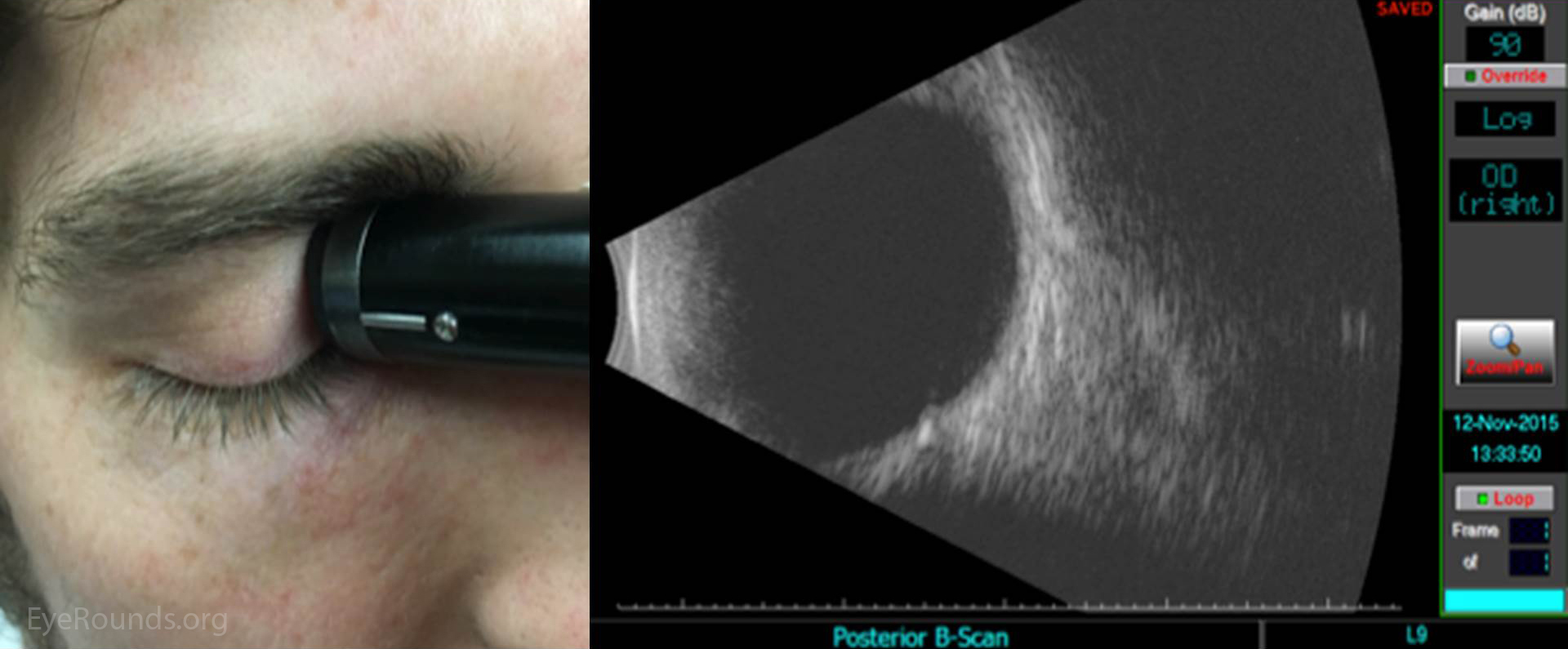
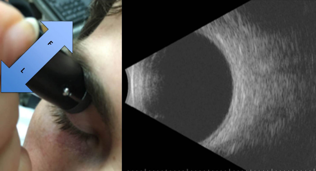
The “b scan” is usually performed by placing the transducer on the patient’s closed eyelid. An a/b scan & ubm ultrasound platform. It is otherwise called brightness scan.
The b scan probe has a marking. Conventionally the marking denotes the upper part of b scan. The orientation of b scan probe may be longitudinal, transverse or axial.
With understanding of the indications for ultrasound and proper examination. It can provide additional information not readily obtained by direct visualization of ocular tissues, and it is particularly useful in patients with pathology that prevents or obscures. The echoes of which are represented as a multitude of dots that together form.
Aviso is used in most of the ultrasound teaching institutions across the world. Use on tabletop or attach to vesa mount. Ultrasound biomicroscopy (ubm) is performed with a much higher frequency than the a and b scans and is primarily used to evaluate the anterior chamber angle and ciliary body.
It is commonly used to see inside the eye when media is hazy due to cataract or any corneal opacity. It creates the typical 2d image that you associate with ultrasound. With an installed base of more than 2500 units worldwide, aviso is the reference in ophthalmic ultrasound imaging thanks to its excellent image quality for posterior pole and anterior segment diagnosis.
The stronger the echo, the higher the spike. A survey of the current status of b scan ultrasonography in ophthalmology, with examples of ultrasound photographs, is presented. The echoes of which are represented as spikes arising from a baseline.
Truly outstanding image quality and elegant user interface. The echographic exam of the human eye and orbit is described, and echographic characteristics of various ocular. Product estimates & trend analysis 4.1.