24 normal reference values for. A z score of +2 or 2 corresponds to the 95th percentile (i.e., 2 standard deviations above or below the mean) nomograms and z scores.

Various formulas can be used to calculate areas, volume, ejection fraction, fractional shortening, etc.
2d echocardiography normal values. ∗∗ in the absence of other etiologies of lv and la dilatation and acute mr. Various formulas can be used to calculate areas, volume, ejection fraction, fractional shortening, etc. Aggarwal s, pettersen md, gurckzynski j et al.
Rature and accepted as normal limits. Relative wall thickness (cm)* = 2 x posterior wall / left ventricular diastolic diameter. 24 normal reference values for.
A z score > 2 indicate dilatation while a z score <2 indicate hypoplasia. Recent data obtained from three cohorts of >2,400 patients now provide normal values of ra dimensions for men and women.12,73,165. The two dimensions presented are width (x axis) and depth (y axis).
Echo/doppler findings in pericardial tamponade Data baserad på linjär regressionsmodell av biaggi et al (gender, age, and body surface area are the major determinants of ascending aorta dimensions in subjects with apparently normal echocardiograms, j am soc echocardiogr. 2d echocardiography can measure areas, circumferences and lengths from the caliper and tracing function on the echocardiographic machine.
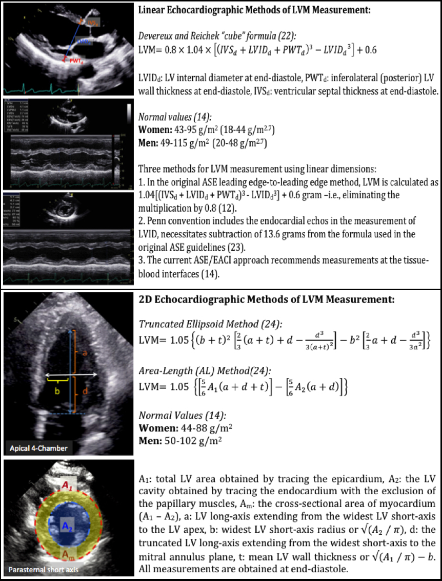
They are based in original studies of individuals who were evaluated for suspected heart disease, which was not confirmed thereafter 2,3. Echocardiogram, normal values the normal values for echocardiographic measurements presented in textbooks 1 are heterogeneous and at times inconsistent. Truncated ellipsoid method or area length method.
The base of the rv free wall provides one of the most visibly obvious movements on normal echocardiography : The norre (normal reference ranges for echocardiography) study is the first european, large, prospective, multicentre study performed in 22 laboratories accredited by the european association of cardiovascular imaging (eacvi) and in 1 american laboratory, which has provided reference values for all 2d echocardiographic (2de) measurements of the 4 cardiac. Results of the world alliance of societies of echocardiography study.
A limitation of the 2d methods is that they rely on geometrical assumptions that are not applicable when there are major lv distortions or when the lv is foreshortened. Normal myocardial strain values using 2d speckle tracking echocardiography in healthy adults aged 20 to 72 years echocardiography. Area can be measured using the echocardiographic tracing function.
Thus to normalize in pediatric echocardiography we use nomograms normalized data are expressed as z score i.e. In contrast to the left atrium, ra size appears to be gender dependent, but prior ase guidelines did not have sufficient data to provide normative data by gender.1,71. After normalization for the body surface area, la volumes were no longer different between groups.
∗∗ in the absence of other etiologies of lv and la dilatation and acute mr. As fluid accumulates in the pericardial space, intrapericardial pressure increases,eventually equaling or exceeding intracardiac pressures. Click here for calculation of lv mass.
A tapse cutoff value < 17 mm yielded high specificity, though low sensitivity to The standard ultrasound transducer for 2d echocardiography is the phased array transducer, which creates a sector shaped ultrasound field (figur 1). A z score of +2 or 2 corresponds to the 95th percentile (i.e., 2 standard deviations above or below the mean) nomograms and z scores.