Eroa = (csav)/v (maxmr) { { flödeshastighet = csav = 6.28r2v (aliasing) references. The z score of a measurement is the number of standard deviations of that value from the mean.

Ejection fraction can be measured with imaging techniques, including:
2d echo normal values. One data set that an echocardiogram provides is ventricular dimensions. Echo is the cheapest and least invasive method available for screening cardiac anatomy. A tapse cutoff value < 17 mm yielded high specificity, though low sensitivity to
The normal values for echocardiographic measurements presented in textbooks 1 are heterogeneous and at times inconsistent. Tmt positive 2d echo normal 4647 views my tmt is positive for inducible ischameia. The approximate normal values for various cardiac structures are described in table 1.
The norre (normal reference ranges for echocardiography) study is the first european, large, prospective, multicentre study performed in 22 laboratories accredited by the european association of cardiovascular imaging (eacvi) and in 1 american laboratory, which has provided reference values for all 2d echocardiographic (2de) measurements of the 4 cardiac. Highly dependent on probe rotation which can result in an underestimation of rv width. • end diastole • diameter > 41 mm (base) and > 35 mm (mid level)=rv dilatation • > 83 mm (longitudinal) = rv enlargement.
Ef indicates to the percentage of blood pumped out of heart chamber during each heat beat (systole). The sampling criteria of the present study differentiate it from most of the studies reported in the literature, because A limitation of the 2d methods is that they rely on geometrical assumptions that are not applicable when there are major lv distortions or when the lv is foreshortened.
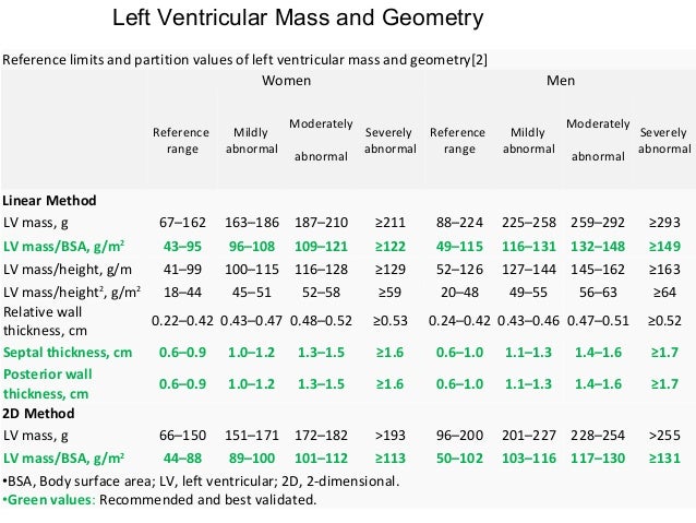
Click here for calculation of lv mass. They are based in original studies of individuals who were evaluated for suspected heart disease, which was not confirmed thereafter 2,3. 24 normal reference values for.
In this study we present the normal reference values of echocardiographic chamber dimensions in young eastern indian adults and compare it with the data in present guidelines and recent studies involving indian subjects. 6 in the present study, we reported the reference ranges for normal doppler parameters taking age and gender into account acquired using recommended echocardiographic. Easily obtained and a marker of rv dilatation.
Standard transthoracic echocardiographies were performed to obtain basic. Aggarwal s, pettersen md, gurckzynski j et al. The z score of a measurement is the number of standard deviations of that value from the mean.
Ejection fraction can be measured with imaging techniques, including: How to express normalized data: The visceral pericardium, which is contiguous with the epicardial surface of the heart, and the parietal pericardium, which is a thin fibrous structure closely apposed to pleural surfaces laterally and that merges with the diaphragm inferiorly.
Truncated ellipsoid method or area length method. The base of the rv free wall provides one of the most visibly obvious movements on normal echocardiography : Rv focused apical 4 chamber view.
Results of the world alliance of societies of echocardiography study. Echocardiography (echo).2,3 the normal pericardium consists of two layers: Z scores normalized data may be expressed in different ways, including the percentage of the mean normal value, percentile charts, and z scores.
A normal lv ejection fraction is 55 to 70 percent. ∗∗in the absence of other etiologies of lv and la dilatation and acute mr. Relative wall thickness (cm)* = 2 x posterior wall / left ventricular diastolic diameter.
Importantly, compared with the previous guidelines, this update is. 1 mm st is seen in lead ii, iii, ang, v4 to v6 during exercis. Generalists most commonly request an echo to assess left ventricular (lv) dysfunction, to rule out the heart as a thromboembolic source, and to characterize murmurs.
Eroa = (csav)/v (maxmr) { { flödeshastighet = csav = 6.28r2v (aliasing) references. After normalization for the body surface area, la volumes were no longer different between groups.